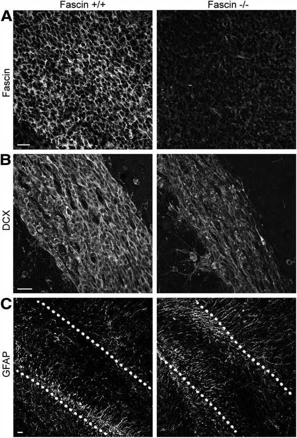
Figure 2.
Fascin-1ko mice show abnormal neuroblast chain organization in the RMS. A, Immunostaining of the SVZ/RMS in sagittal brain slices from P7 mice showing the absence of fascin in fascin-1ko animals. B, Dcx-positive neuroblast chains appear thinner in fascin-1ko mice compared with wt mice. C, Lack of fascin does not appear to perturb the localization of GFAP+ astrocytes and stem cells. The dotted lines outline the RMS borders. Scale bars: A, B, 20 μm; C, 50 μm.

