Figure 9.

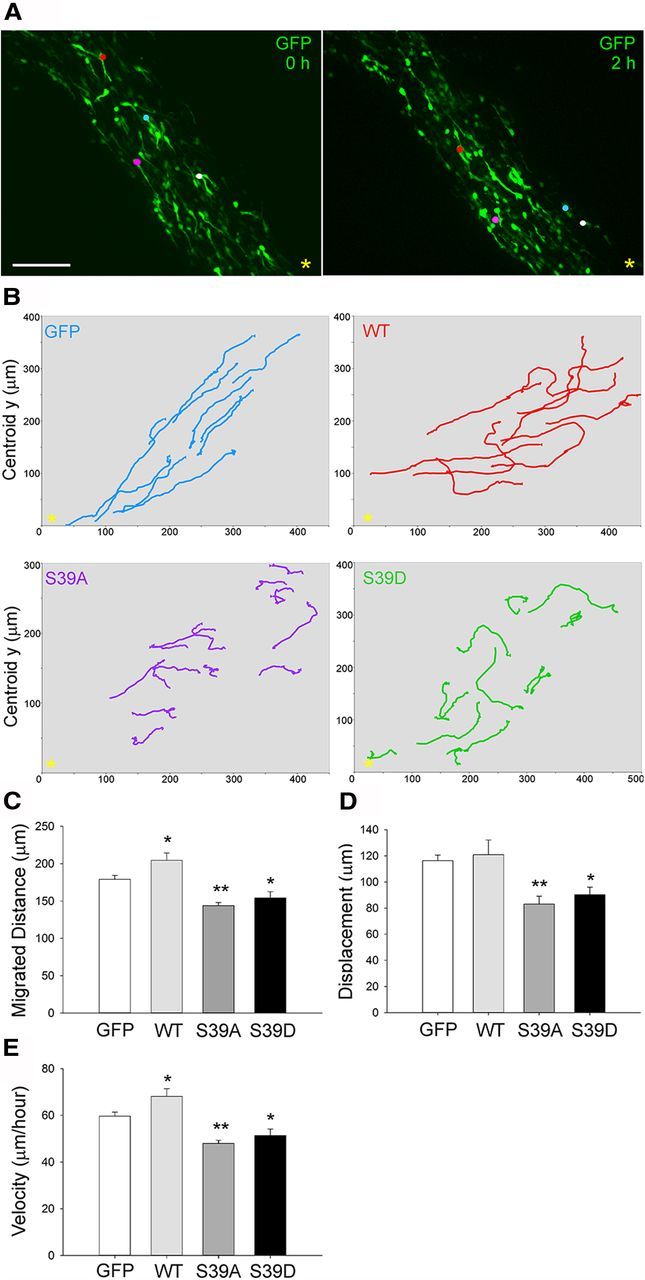
Phosphorylation of fascin on Ser39 regulates neuroblast migration ex vivo. A, P2 mice were electroporated in the lateral ventricle with pGFPC2 (empty vector); pGFPC2-wt, -S39D, or -S39A fascin; and pCX-EGFP in a 3:1 ratio. Acute brain slices were prepared 5 d after electroporation. Projections of spinning disk confocal z-stack images (taken at times 0 and 2 h) from time-lapse microscopy showing GFP-expressing neuroblasts (colored dots) migrating toward the OB (yellow asterisk). B, Neuroblasts expressing wt, S39A, or S39D fascin tend to display an enhanced exploratory behavior compared with control cells expressing only GFP, as shown here by some representative migratory paths from time-lapse imaging of brain slices over a period of 3 h. The yellow asterisk marks the location of the OB. C–E, Based on quantitative tracking analysis, the expression of S39D or S39A fascin significantly decreases migrated distance, speed, and displacement, while wt fascin overexpression increases migration distance and speed (mean ± SEM; n = 8 slices for control; n = 5 slices for wt; and n = 6 slices for S39A and S39D; *p < 0.05; **p < 0.01). Scale bar, 85 μm.
