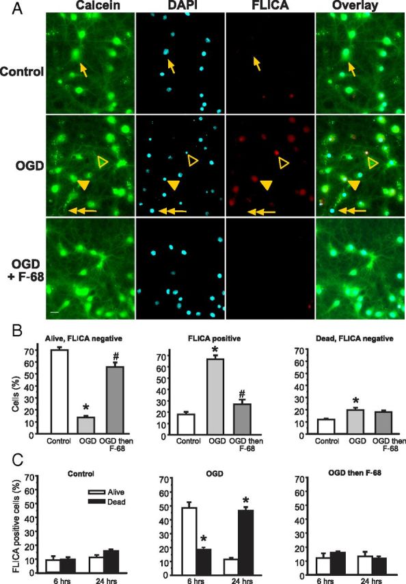
Figure 3.

F-68 prevents OGD-induced caspase activation. A, Using calcein (green) to label living neurons, DAPI (cyan) to label all neurons, and FLICA (red) to label caspase-positive neurons allows identification of neurons as living/dead and caspase activation as positive/negative. Images obtained 6 h after 45 min exposure to control buffer or OGD, with and without immediate treatment thereafter with F-68 (30 μm). Scale bar, 10 μm. Arrow, Living neuron, FLICA negative; filled arrowhead, living neuron, FLICA positive; open arrowhead, dead neuron, FLICA positive; double arrowhead, dead neuron, FLICA negative. B, C, Each experimental unit (n) consists of six coverslips per condition, with high-throughput imaging counting ∼1300 cells per coverslip. * indicates significantly different from control; # indicates significantly different from OGD. B, Mean ± SD percentage of total cells at 6 h after OGD or control (n = 3). C, Mean ± SD percentage of FLICA-positive cells over time after OGD or control (n as in B). * indicates significantly different from the paired bar.
