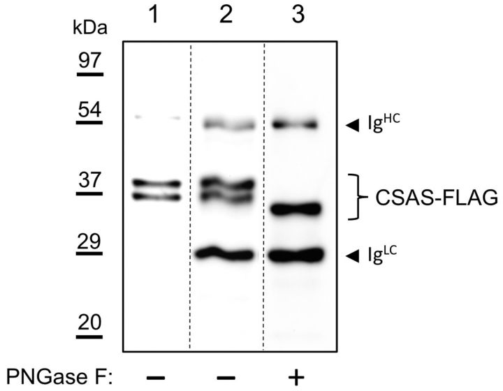
Figure 3.
CSAS protein is modified with N-linked glycans when expressed in vivo. Western-blot analysis of CSAS-FLAG expressed in Drosophila heads. Lane 1, Cell lysate from heads. Lane 2, Affinity purified CSAS-FLAG, PNGase F mock-treated control. Lane 3, Affinity purified CSAS-FLAG treated with PNGase F. The decrease of CSAS molecular mass upon PNGase treatment indicates the presence of N-linked glycans. IgHC and IgLC indicate Ig chains leached from FLAG-affinity beads in denaturing conditions. Approximate positions of molecular mass markers are shown on the left.

