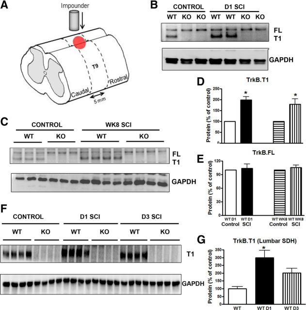
Figure 1.
The trkB.T1 protein is significantly upregulated at the thoracic injured area and lumbar spinal cord horn after SCI. A, Schematic drawing of the area of injury and region of tissue harvested. A moderate contusion injury was produced using a spinal cord impactor with a force of 60 kdyn. Five millimeters of spinal cord tissue centered on the epicenter or equivalent tissue from a laminectomy control was processed for Western blot and microarray analysis. B–C, Representative Western blot showing trkB.FL and trkB.T1 in the intact spinal cord (control), at 1 d and 8 weeks after SCI (D1 and WK8 SCI) in trkB.T1+/+ and trkB.T1−/− mice. D–E, In the trkB.T1+/+ mice, trkB.T1 protein expression was significantly upregulated 1 d after SCI (n = 4) compared with control (n = 4). The upregulation was sustained at week 8 (n = 8 per group, p < 0.05 SCI vs control, two-tailed Student's t test). There was no difference in the expression of the trkB.FL protein at 1 d after SCI or at week 8 after SCI compared with control (n = 4 per group). F, Representative Western blot showing trkB.T1 expression at the lumbar SDH in the intact spinal cord (control) at 1 and 3 d after SCI (D1 and D3 SCI) in trkB.T1+/+ and trkB.T1−/− mice. G, TrkB.T1 expression in TrkB.T1+/+ mice was significantly upregulated at 24 h after thoracic injury (p < 0.05, D1 SCI vs control) and remained elevated at 3 d (n = 4 mice per group). Data are expressed as mean ± SEM.

