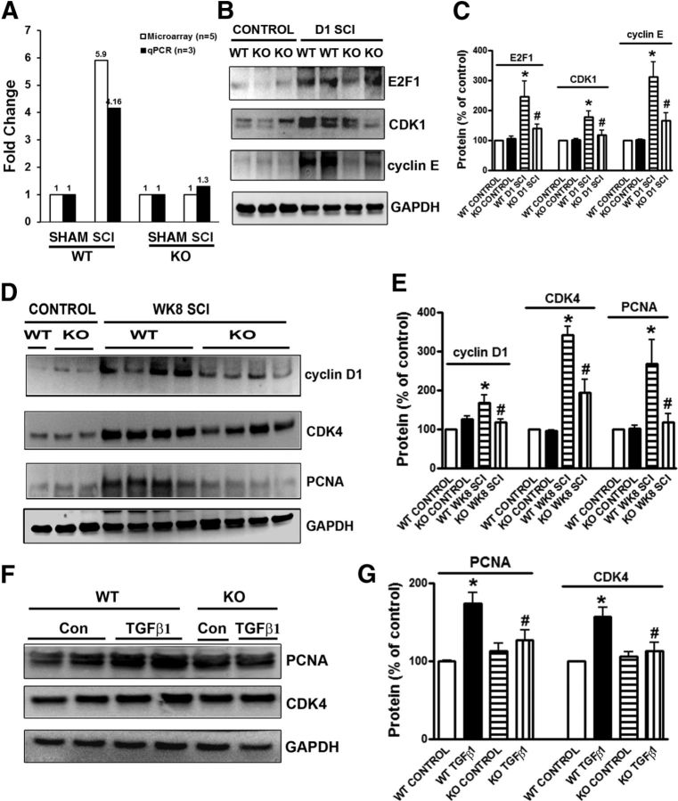
Figure 6.
Deleting trkB.T1 attenuates upregulation of cell-cycle-related proteins in the lesioned and lumbar areas of post-SCI spinal cord and reactive astrocytes in vitro. A, Cdk1 is significantly upregulated 24 h after SCI in trkB.T1+/+ spinal cord by microarray (n = 4, 5.9 FC, FDR p = 2.09E-10) and qPCR (n = 4, FC 4.16, p = 0.0022), but not in trkB.T1−/− mice (n = 4). B, Representative immunoblots of E2F1, CDK1, and cyclin E in spinal cord tissue from trkB.T1+/+ and trkB.T1−/− mice 24 h after SCI. C, Quantification showing E2F1, CDK1, and cyclin E protein upregulation in trkB.T1+/+ and trkB.T1−/− mice compared with controls, which was significantly attenuated in trkB.T1−/− compared with trkB.T1+/+ mice (n = 4 mice/group). D, Representative immunoblots of cyclin D1, CDK4, and PCNA in spinal cord tissue from trkB.T1+/+ and trkB.T1−/− mice 8 weeks after SCI. E, Quantification showing upregulation of all three proteins in trkB.T1+/+ and CDK4 and PCNA in trkB.T1−/− mice compared with the controls, which was significantly attenuated in trkB.T1−/− compared with trkB.T1+/+ mice (n = 8 mice/group). *p < 0.05, WT SCI versus WT control; #p < 0.05, KO SCI versus WT SCI. F, Representative immunoblots of PCNA and CDK4 in primary astrocyte cultures from trkB.T1+/+ and trkB.T1−/− mice ± TGFβ1. G, Quantification showed significant attenuation of protein upregulation in reactive astrocytes from trkB.T1−/− compared with trkB.T1+/+ mice. *p < 0.05, WT TGFβ1 versus WT control; #p < 0.05, KO TGFβ1 versus WT-TGFβ1; n = 4 independent cultures/group.

