Figure 1.
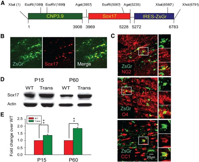
Generation of CNPase promoter-driven Sox17 transgenic mice leads to elevated levels of Sox17 in postnatal WM tissue. A, Diagram showing transgenic construct containing full-length mouse Sox17 cDNA fragment between CNPase promoter and IRES-EGFP. B, Immunohistochemical analysis of total Sox17-expressing cells in the CNP-Sox17 P30 subcortical WM. Confocal microscope images showing colocalization of ZsGreen fluorescence with Sox17 in WM, indicating expression of the transgene. C, Confocal images showing colocalization of ZsGreen fluorescence with NG2 (red), O4 (red), and CC1 (red) in subcortical WM at P30. Scale bar, 20 μm. D, Western blot showing total Sox17 protein levels in corpus callosum (CC) in the CNP-Sox17 transgenic (Trans) compared with WT mice. E, Quantitation of Western blots including those in D. Densitometric values of Sox17 protein normalized against actin are plotted as a function of postnatal age. Values represent the mean fold change of Sox17 protein abundance, relative to actin, in transgenic (Trans) over WT. Increased levels of Sox17 are observed in the CNP-Sox17 (Trans) at P15 and P60. Values shown are mean and SEM obtained from 1–2 mice from each of four independent litters. **p < 0.01 versus WT, unpaired Student's t test.

