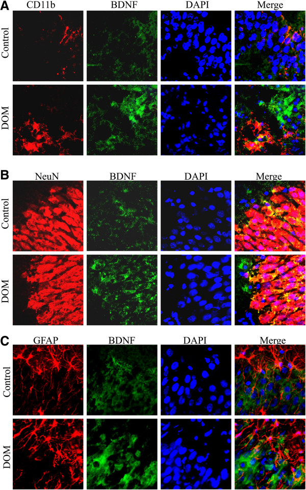
Figure 2.
Immunohistochemical visualization of BDNF in microglia, neurons and astroglial cells in CA1 area, following excitotoxic insult. (A) Representative fluorescence photomicrographs of CD11b-positive microglial cells (red), BDNF (green), DAPI and merge images. Upper row: control culture, in which BDNF-positive microglial cells have resting-like morphology. Lower row: culture exposed to DOM for 24 h and then transferred to a DOM-free medium for 7 days showed highly activated and BDNF-expressing microglia (lower left quadrant). (B) Representative fluorescence photomicrographs of NeuN-positive neurons (red), BDNF (green), DAPI and merge images. Most BDNF immunoreactivity in both control and DOM-treated group was observed in NeuN-positive cells. (C) Representative photomicrographs of GFAP-positive astroglial cells (red), BDNF (green), DAPI and merge images. No significant changes were observed in the number of astrocytes expressing BDNF in presence or absence of the DOM insult.

