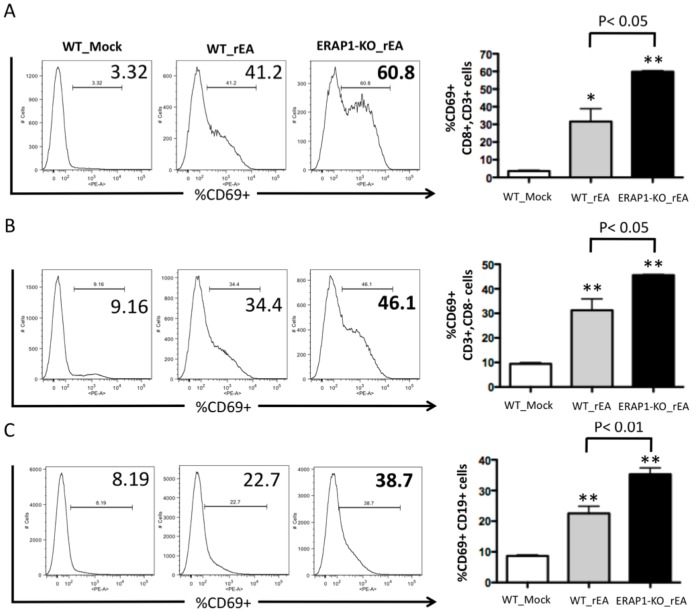
Figure 3. Mice lacking ERAP1 exhibit dramatically enhanced activation of B cells, CD8+ and CD8− T cells in response to rEA stimuli.
C57BL/6 WT and ERAP1-KO mice were either mock (PBS) injected or intraperitoneally injected with 100 ng/mouse of rEA protein. Splenocytes were harvested at 6 hpi, processed, stained for expression of surface markers, and analyzed by FACS as described in Materials and Methods. CD69 activation of (A) CD8+ CD3+ T cells, (B) CD8− CD3+ T cells, and (C) B cells are shown. Bars represent mean ± SEM. Representative plots are shown. Statistical analysis was completed using a one-way ANOVA with a Student-Newman-Keuls post-hoc test. n = 4 for all groups of mice. *, ** - indicate values, statistically different from those in mock-injected mice, p<0.05, p<0.001, respectively.

