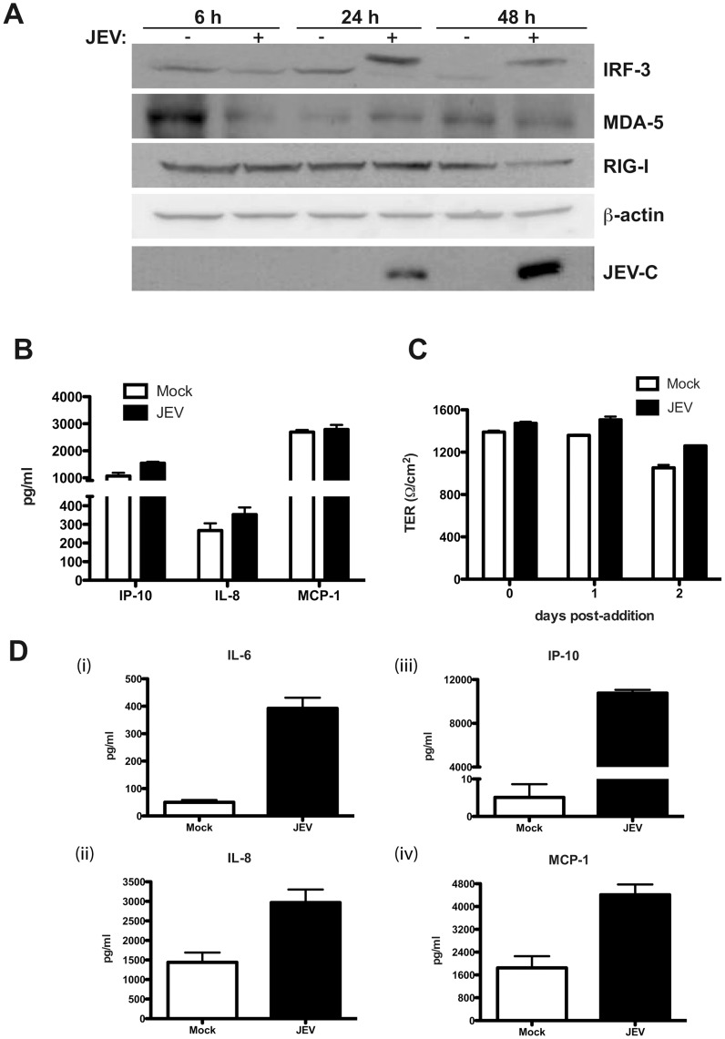
Figure 6. JEV mediated disruption of tight junctions is independent of secreted factors.
(A) Caco-2 cells were infected with JEV and cells were collected at indicated time points for preparation of cell lysates. Induction/activation of the indicated proteins was detected by western blot analysis. β-actin serves as loading control and JEV infection is indicated by the expression of capsid protein (JEV-C). (B) The amount of indicated cytokines in infected culture supernatants were measured by Luminex bead assays as described in materials and methods. The figures are representative of three experiments performed with two or more replicates. Error bars indicate mean ± s.d. (C) Clarified supernatants from JEV-infected cells were added onto naïve caco-2 cells grown on trans-wells and TER levels were monitored for indicated periods. The figures are representative of three experiments performed with two or more replicates. Error bars indicate mean ± s.d. (D) The amount of indicated cytokines in infected culture supernatants of HUVECs were measured by Luminex bead assays at 48 h pi as described in materials and methods. Error bars indicate mean with SEM of three replicates.

