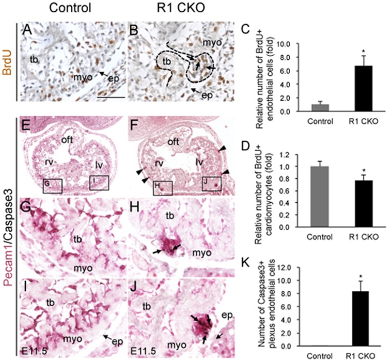
Figure 3. The coronary plexuses in the R1 CKO hearts are unstable and self-limited through apoptosis.
A, B, Images of E11.5 ventricle immunostained with antibodies against BrdU (brown nuclear staining) showing the proliferating plexus endothelial cells in the R1 CKO hearts (B, indicated by arrows and dash line). No plexus is present in the same region of the control heart (G). Scale bar = 50 µm. C, D, Statistical analyses showing a significantly increased proliferation of the plexus endothelial cells (C) and decreased proliferation of cardiomyocytes (D) in the E11.5 R1 CKO hearts. n = 3 individual hearts, 5 comparable sections per heart, error bars = SD. E-J, Images of the frontal sections of E11.5 ventricles co-immunostained with the antibodies against Pecam1 (purple membrane staining) and Caspase3 (black nuclear staining) showing the apoptotic endothelial cells within the overgrowing coronary plexuses in the R1 CKO embryos (F, arrowheads; H and J, arrows). No apoptosis is present in the control hearts (E, G, I). K, Quantitative analysis showing a significantly increased apoptosis of the R1 CKO endothelial cells. n = 3 individual hearts, 5 comparable sections per heart, error bars = SD.

