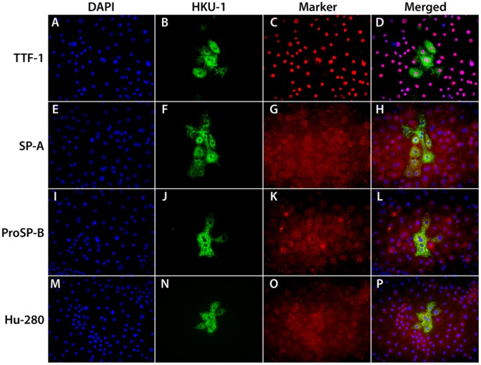
Figure 1. Immunofluorescent staining for HCoV-HKU1 spike protein and selected alveolar type II cell markers.
The cells were grown under air/liquid conditions as described in the methods section, inoculated with HCoV-HKU1 and fixed 72 hours post inoculation. Panels A-D show staining for DAPI (A), HCoV-HKU1 (B), TTF-1 (C), and merged (D). Panels E-H show staining for pro DAPI (E), HCoV-HKU1 (F), SP-A (G), and merged (H). Panels I-L show staining for DAPI (I), HCoV-HKU1 (J), proSP-B (K), and merged (L). Panels M-P show staining for DAPI (M), HCoV-HKU1 (N), AT280 (Dobbs) (O), and merged (P). Cells that are infected with HCoV-HKU1 stain for the type II cell markers.

