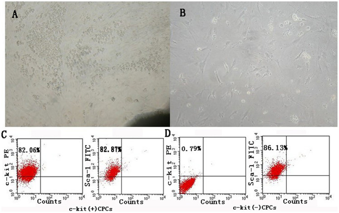
Figure 1. Characterization of cultured CPCs.
(A) Cells (small, round, and phase-bright) migrated from the cardiac explants, and aggregated and proliferated on the fibroblast layer after 10 days of culture (×100 magnification). (B) Representative clone generated by CPCs (×100 magnification). (C) and (D) Representative flow cytometric analyses of c-kit(+)CPCs and c-kit(−)CPCs for the expression of the cell surface markers, namely, c-kit, and Sca-1. The Figure 1 panels A and B are excluded from this article's CC-BY license. See the accompanying retraction notice for more information.

