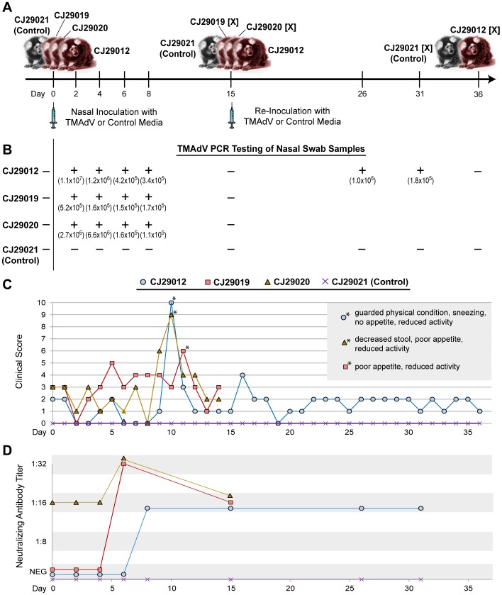
Figure 2. Experimental TMAdV Infection in the Common Marmoset.
(A) Outline of TMAdV infection protocol and sample collections. The “[X]” refers to a marmoset that is sacrificed at the designated timepoint. (B) Results from PCR analysis of nasal swab samples collected at serial timepoints. Shown in parentheses are the calculated viral titers in genome copy numbers per mL associated with each timepoint. (C) Clinical scores in TMAdV-infected and control marmosets. The asterisks (*) refer to timepoints during which each infected marmoset exhibited the most pronounced clinical signs of illness (inset box). For a definition of the clinical scoring system, see Table S2. (D) Antibody titers in TMAdV-infected and control marmosets as measured using a TMAdV neutralization assay.

