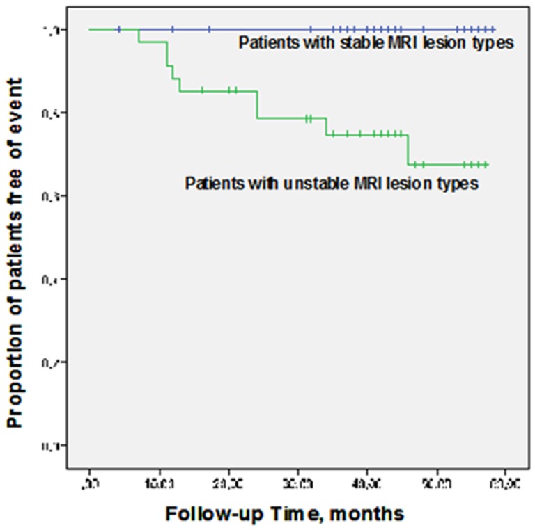
Figure 3. Kaplan–Meier curves.

Kaplan–Meier survival estimates of the proportion of patients free of ipsilateral cerebrovascular events for patients presenting with stable MRI lesion types (upper curve) and with unstable MRI lesion types (lower curve). Event-free survival was higher among patients with the MRI-defined stable lesion types (III, VII, and VIII) than in patients with the MRI-defined high-risk lesion types (IV–V and VI) (log rank test P<0.0001).
