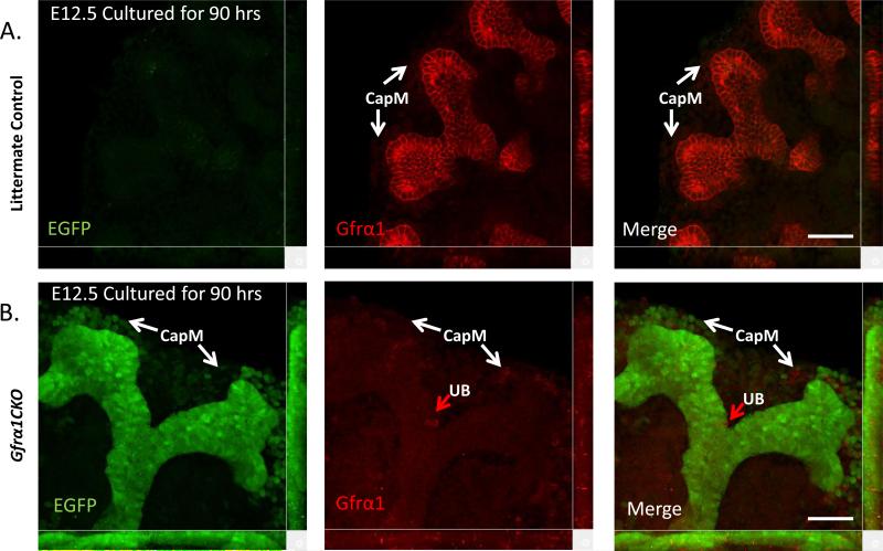
Figure 6. Robust excision of Gfrα1 in UB epithelium metanepheroi cultured ex-vivo.
Confocal microscopy images obtained from EGFP (green) and Gfrα1 (red) double immunofluorescence labeling of control (A) or Gfrα1CKO (B) kidneys cultured for 90 hours in presence of 4HT. Note robust green signal and only a few Gfrα1-labeled (red arrow) cells in Gfrα1CKO panels indicate highly efficient Gfrα1 excision in the UB tip. There is only partial excision of Gfrα1 in the cap mesenchyme (CapM). Scale bar 50 μm.

