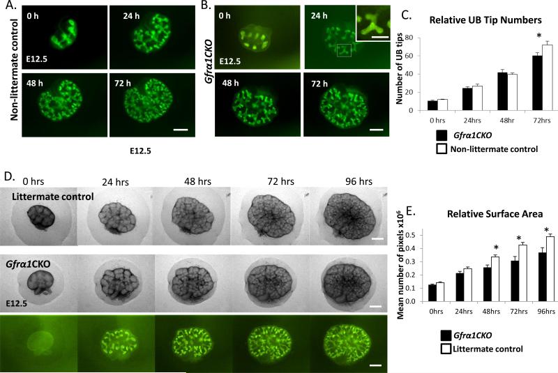
Figure 7. Metanephric kidney culture experiments show a modest decrease in branching in Gfrα1CKO mice.
(A-B) Fluorescent images of metanephroi from (A) control (Gfrα1EGFP/+) and (B) Gfrα1CKO embryos dissected at E12.5 and cultured in presence of 4HT at the indicated time points in hours (h). To ensure controls and Gfrα1CKO mice had similar number of UB tips at the beginning of the culture pregnant mothers were treated with 4HT the night before sacrifice to induce EGFP expression. Inset in B at 24h is higher magnification of the area depicted by white square and it shows presence of EGFP positive cells in almost the entire branching UB tip indicating near complete excision. Note Gfrα1CKO kidneys continue to branch and green cells colonize the tips. (C) Bar graph shows comparison between the number of UB tips in cultured metanephroi of Gfrα1CKO mice and controls at indicated time points. A mild reduction in branching is seen at 72 hours. Results are represented as mean±s.e.m (Control n=11, Gfrα1CKO n=9. *P<0.05 by t-test). (D) Phase images of littermate control (Gfrα1floxEGFP/+) metanephroi compared to phase and fluorescent images of Gfrα1CKO dissected at E12.5, cultured in the presence of 4HT. (E) Bar graph shows comparison between surface areas (mean number of pixels per metanephros ±s.e.m; *P<0.01 by t-test) of Gfrα1CKO (n= 7) and littermate controls (n=12) at indicated time points. Scale bar: 250 μm (inset scale bar 125 μm).

