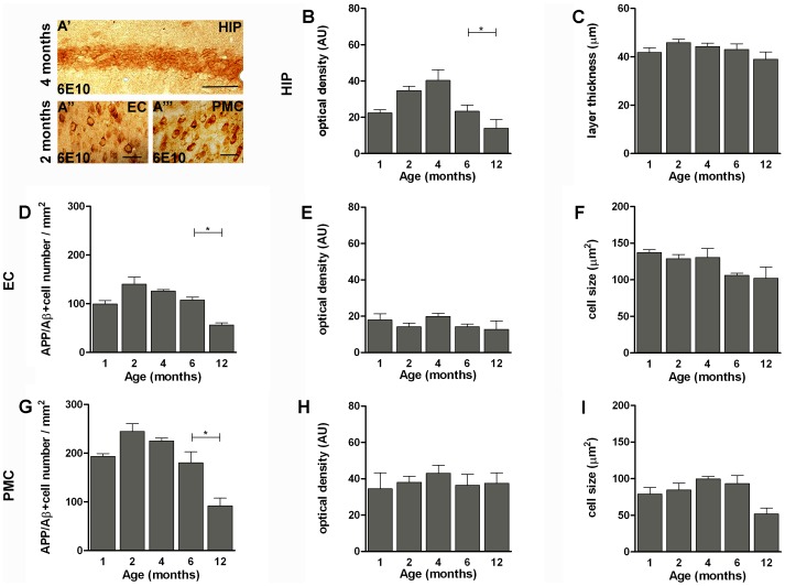
Figure 2. Time course of intracellular APP/Aβ immunoreactivity in APPSwe/PS1L166P mice.
Representative images of coronal sections of hippocampal pyramidal cell layer (A′), entorhinal (A′′) and primary motor (A′′′) cortex of APPSwe/PS1L166P mice, at the peak of intracellular 6E10 immunostaining positivity. Calibration bar, 50 µm. (B-I) Quantitative analysis of APP/Aβ immunoreactivity in the intracellular compartment in hippocampal pyramidal cell layer (B; C), entorhinal (D; E; F) and primary motor cortex (G; H; I) in terms of APP/Aβ+ optical density (B; E; H), hippocampal pyramidal cell layer thickness (C), cell number (D; G) and cell size (F; I) in 1-, 2-, 4-, 6- and 12-month-old APPSwe/PS1L166P mice. The APP/Aβ+ optical density of the intracellular compartment in hippocampal pyramidal cell layer (B) and the APP/Aβ+ cell number in both entorhinal (D) and primary motor (G) cortex showed age-related differences. The hippocampal pyramidal cell layer thickness (C) and the APP/Aβ+ cell size in both entorhinal (F) and primary motor cortex (I) showed no age-related differences (n = 5 in each group, three sections per mouse, three frames per section, * P<0.05, One-way ANOVA). Error bars represent SEM. (AU, arbitrary units; HIP, hippocampus; EC, entorhinal cortex; PMC, primary motor cortex).

