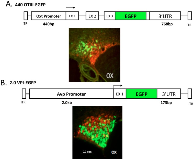
Figure 1. Specificity of Expression of the Oxytocin and vasopressin rAAV constructs in the SON.
A. The 440OTIII-EGFP rAAV that was injected into the SON contains 440 bp of the promoter sequence upstream of the transcription start site (TSS) of the oxytocin gene, all three exons, fused to the EGFP sequence within the 3rd exon (EX 3), followed by 768 bp of the 3′UTR of the oxytocin gene [17]. The immunohistochemical (IHC) image shown below the construct shows that the EGFP from the 440 OTIII-EGFP virus is specifically expressed in Oxt MCNs in the dorsal SON. The green color represents the fluorescence of the EGFP immunoreactivity in Oxt MCNs and the red fluorescence depicts the PS 41 antibody immunoreactivity in the Avp MCNs. Scale line for both panels A and B is shown in lower panel B. B. The 2.0VPI-EGFP rAAV that was injected contains 2.0 bp of the promoter sequence found upstream of the vasopressin gene TSS, exon 1 of the vasopressin gene, fused to the EGFP sequence, and 173 bp of the 3′UTR downstream of the Avp gene [18]. The immunohistochemical (IHC) image shown below the construct shows that the EGFP from the 2.0VPI-EGFP virus is specifically expressed in Avp MCNs in the ventral SON. The green color represents the fluorescence of the EGFP immunoreactivity in Avp MCNs and the red fluorescence depicts the PS 38 antibody immunoreactivity in the Oxt MCNs. Abbreviation: OX, optic chiasm.

