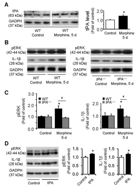
Figure 4. Chronic morphine induces IL-1β and pERK expression in astrocyte cultures via tPA.
(A) Western blot showing tPA expression following chronic morphine treatment (100 μM, 5 days) in astrocyte cultures from WT mice. The line under the gel indicates the two parts are from the same gel but not adjacent. Right panel, intensity of tPA bands. *P<0.05, compared to Control (saline), t-test, n=4 cultures. (B) Western blots showing pERK and IL-1β expression in astrocytes from WT (left blot) and tPA−/− (right blot) mice before and after morphine treatment. (C) Intensity of pERK (p42/44) and IL-1β bands in astrocytes from WT (left graph) and tPA−/− (right graph) mice before and after morphine treatment. Morphine increases pERK and IL-1β expression in wild-type but not tPA-deficient astrocytes. *P<0.05, compared to control; #P<0.05; n=4 cultures. (D) tPA (50 ng/ml, 1 day) induces pERK and IL-1β expression in WT astrocytes. *P<0.05, compared with control, n=4 cultures.

