Figure 1.
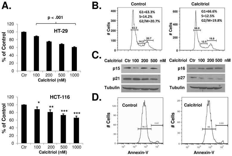
Calcitriol inhibits the in vitro growth of colon cancer cells. (A) HT-29 and HCT-116 cells were treated with various doses of calcitriol for 48h and in vitro cell growth was analyzed using WST-1 assay as described in Materials and Methods. (B) HCT-116 cells were treated with 500 nM calcitriol for 48h and cell cycle distribution was analyzed by flow cytometry. The experiment was repeated three times; representative results of three independent experiments were shown. The percentages of cells in each phase of cell cycle (mean ± S.D.) are: Control: G1 (60.6 ± 3.2), S (14.5 ± 0.4), G2 (22.5 ± 1.9); Calcitriol: G1 (65.0 ± 2.7), S (12.8 ± 0.4), G2 (21.2 ± 2.0). (C) HCT-116 cells were treated with various doses of calcitriol for 48h and the expression of cell cycle regulatory proteins was analyzed by western blots. The experiment was repeated three times. Representative images of three independent experiments were shown. (D) HCT-116 cells were treated with 500 nM of calcitriol for 48 hours, apoptosis was determined by annexin V staining and flow cytometry. The experiments have been repeated three times, representative results of three independent experiments were shown. Data shown are mean values + SD (*p<0.05, **p<0.01, ***p<0.001).

