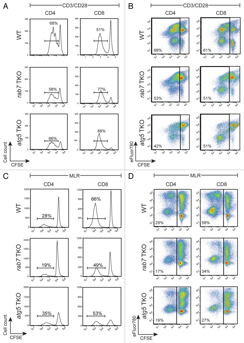
Figure 7. Deletion of rab7 or atg5 produced similar defects in activated T cells. (A) Freshly purified, CFSE-labeled splenic T cells were activated with plate-bound anti-CD3 and soluble anti-CD28 antibodies. At 65 h, cells were surface-stained for CD4 and CD8 and then incubated with eFluor780 prior to analysis by flow cytometry. CFSE levels in live-gated CD4+ or CD8+ populations are shown and the percent of live cells that diluted CFSE is indicated. (B) As in (A), but without live gating; percentage of all cells that proliferated (diluted CFSE) is shown. (C and D) CFSE-labeled purified T cells isolated from age- and sex-matched wild-type (WT), rab7 TKO, and atg5 TKO mice were mixed with mitomycin C-treated splenocytes from a Balb/c mouse. After 84 h of incubation, cells were surface-stained for CD4 and CD8, labeled with eFluor780, and CFSE dilution evaluated in the H-2Dd-negative (C57BL/6) population. Live (C) or total (D) cells are shown; percentages indicate the fraction of cells that diluted CFSE. In (A–D), a representative experiment is shown; these experiments were repeated at least 3 times with similar results.

An official website of the United States government
Here's how you know
Official websites use .gov
A
.gov website belongs to an official
government organization in the United States.
Secure .gov websites use HTTPS
A lock (
) or https:// means you've safely
connected to the .gov website. Share sensitive
information only on official, secure websites.
