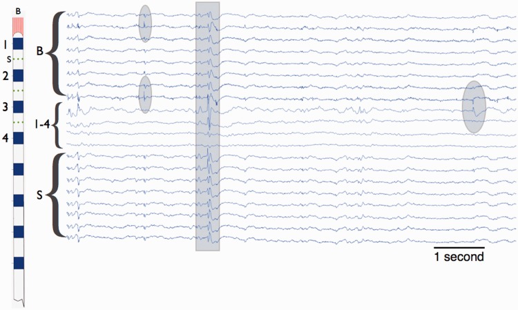
Figure 2.
Intracranial EEG recorded from mesial temporal lobe with a hybrid depth electrode. Left: Hybrid depth electrode with bundle of eight micro-electrodes exiting the tip (red, labelled B), eight clinical macro-electrodes (blue, labelled contacts 1–4), and nine microelectrodes along the depth shaft (red, labelled S). Right: Eight seconds of data from wake patient. From the top, channels 1–7 (labelled B) are from the microwires extending from the electrode tip, channels 8–11 are from the clinical macroelectrodes (labelled 1–4), and the channels 16–20 are from the shaft microwires. The ovals highlight focal interictal epileptiform discharges that are localized to single channels, and the rectangle highlights a diffuse interictal epileptiform discharges occurring on multiple micro and macroelectrode channels.

