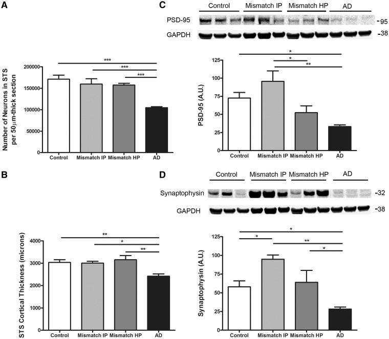
Figure 2.
Number of neurons, cortical thickness and synaptic markers in the superior temporal sulcus. (A) Stereologically based neuronal counts in the superior temporal sulcus (STS) showed a significant reduction by >40% in the number of neurons in demented cases with Alzheimer’s disease (AD) compared to controls. Intermediate (Mismatch IP) and high probability mismatches (Mismatch HP) showed no significant neuronal loss in this region compared with controls. (B) Superior temporal sulcus cortical thickness was significantly reduced by >20% in cases with Alzheimer’s disease but not in intermediate or high probability mismatches when compared to controls. (C) Representative image of western blot and quantification for postsynaptic marker PSD-95. Levels of PSD-95 in the superior temporal sulcus were significantly decreased in cases with Alzheimer’s disease but not in intermediate or high probability mismatches when compared with controls. (D) Representative image of western blot and quantification for presynaptic marker synaptophysin. Levels of synaptophysin in the superior temporal sulcus were significantly decreased in cases with Alzheimer’s disease but not in high probability mismatches when compared with controls. A significantly higher level of synaptophysin was detected in the superior temporal sulcus in intermediate probability mismatches compared with controls. GAPDH was used as loading control. n = 5–8 per group; *P < 0.05; **P < 0.01; ***P < 0.001. One way ANOVA and post hoc Tukey test.

