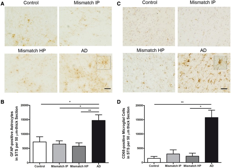
Figure 8.
Number of GFAP-positive astrocytes in the superior temporal sulcus and CD68-positive microglia. (A) Representative photomicrographs of GFAP-positive astrocytes. (B) Stereological counts of astrocytes in the superior temporal sulcus (STS) on sections immunostained with GFAP showed a significant increase in the amount of GFAP-positive astrocytes in demented cases with Alzheimer’s disease but not in intermediate probability (Mismatch IP) or high probability mismatches (Mismatch HP) in comparison with controls. (C) Representative photomicrographs of CD68-positive microglia cells in the superior temporal sulcus. Haematoxylin was used as a counterstain. (D) Stereological counts of microglia in the superior temporal sulcus on sections immunostained with CD68 showed a significant increase in the amount of CD68-positive microglial cells in demented cases with Alzheimer’s disease, but not in intermediate or high probability mismatches in comparison with controls. n = 8–14 per group; *P < 0.05; **P < 0.01. One way ANOVA and post hoc Tukey test, and Kruskal-Wallis ANOVA and Dunn’s multiple comparison test, respectively. Scale bar = 100 µm.

