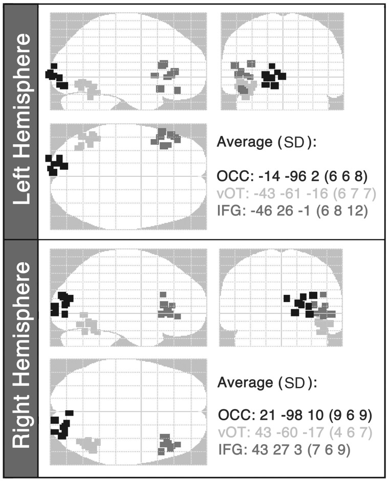
Figure 2.
Optimal source locations from the variational Bayesian equivalent current dipole analysis for each subject plotted on a glass brain in MNI space. The starting points for the source locations were: occipital cortex: ±15 −95 2; ventral occipitotemporal cortex: ±44 −58 −15 14; inferior frontal gyrus: ±48 28 0. The average locations (with standard deviations, SD) of the winning source locations for the patient group are also reported. OCC = occipital cortex; vOT = ventral occipitotemporal cortex; IFG = inferior frontal gyrus (n = 8).

