
Figure 2.
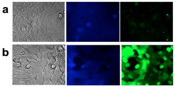
HEK and DMT-1 cells treated with: 5 μM Mn2+, 3.6 μM Fz1AM, 26.6 μM CB-AM and 10 μM Cd2+. (a) HEK cells show blue fluorescence but little green fluorescence, suggesting minimal Mn2+ present in the cell. (b) DMT-1 cells under the same conditions show lower blue fluorescence with bright green fluorescence indicative of markedly increased Mn2+ uptake.