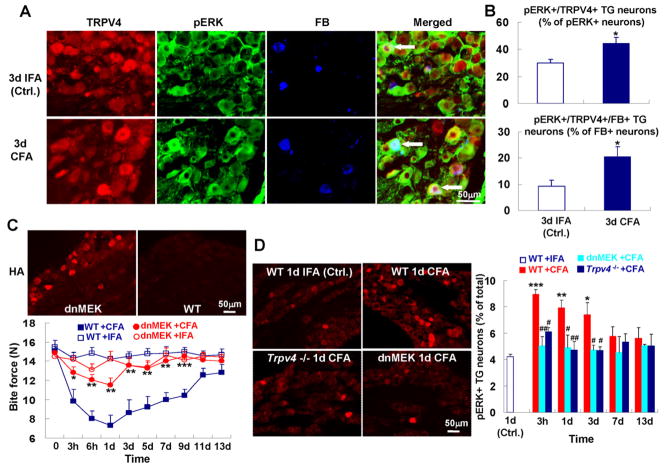
Fig. 6.
MAP-kinase signaling down-stream of TRPV4 is critical for reduction of bite force in TMJ inflammation. (A) pERK-TRPV4 co-expressing TG neurons innervate the TMJ. (B) pERK expressing TG neurons that innervate the TMJ and co-express TRPV4. The upper panel depicts the increase for pERK-TRPV4 co-expressing neurons, of all pERK+ TG neurons, the lower panel the increase for TMJ-innervating pERK-TRPV4 co-expressing neurons, of all TMJ-innervating neurons (n=5 animals per group; *p<0.05; two-tail t test). (C) ERK phosphorylation in neurons is required for reduction of bite force in response to TMJ inflammation. Micrographs: note presence of the transgene, identified by HA-immunolabeling in the TG of a dnMEK-transgenic mouse. Lower panel: Note reversal of attenuation of bite force for dn-MEK-transgenic mice to levels as seen in Trpv4−/− mice (n=7–9 mice per group; *p<0.05, **p<0.01, ***p<0.001; one-way ANOVA with Tukey post test). (D) Increase of pERK expressing TG neurons in response to TMJ inflammation depends on Trpv4. Left-hand micrographs show pERK expression in the TG, revealed by immunolabeling. Right-hand bar graphs depict quantitation. Note early and robust increase of pERK-expression in the TG, and virtually complete lack thereof in Trpv4−/− mice; also note similar lack in dn-MEK-transgenic mice, as expected (n=4–5 mice per group; *p<0.05, **p<0.01, ***p<0.001, WT+CFA vs. WT+IFA; #p<0.05, ##p<0.01 Trpv4−/− or dn-MEK-transgenic mice vs. WT, all + CFA; two-tail t test).

