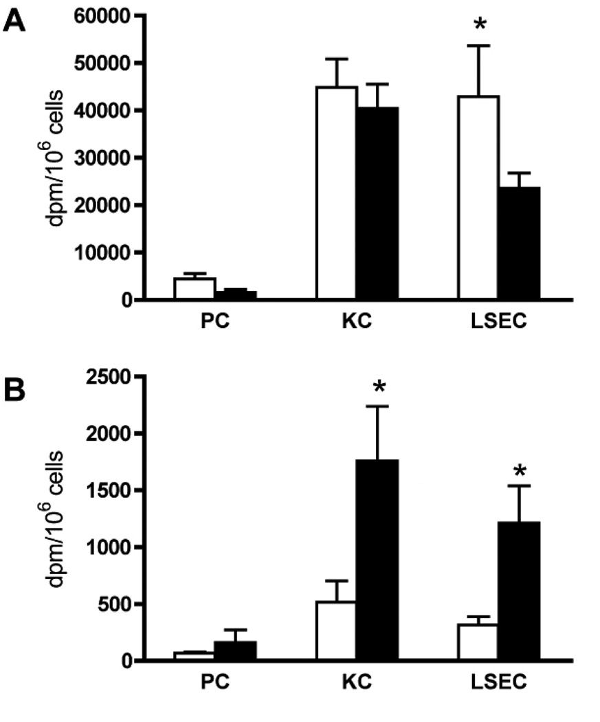
Figure 3. Hepatocellular distribution of 14C-AGEs in pre-pubertal and young-adult mice.
Pre-pubertal and young-adult mice were injected intravenously with 14C-AGE-BSA. The animals were euthanized after 10 min (A) or 4 weeks (B) and radioactivity (dpm/million cells) was measured in solubilized cultures of purified parenchymal cells (PC), Kupffer cells (KC), and liver sinusoidal endothelial cells (LSECs). AGE-BSA injection dose: 9.4 µg/animal; in the 10 min group this dose was achieved by mixing 2.35 µg 14C-AGE-BSA (1×10⁶ dpm) with 7.05 µg non-radioactive AGE-BSA, while the 4 week group was injected with 9.4 µg of 14C-AGE-BSA (4×10⁶ dpm).
A) Radioactivity# in liver cells 10 min after ligand injection in 0.8 month old (open bars, n=7) and 3 month old mice (filled bars, n=6). #Adjusted values.
B) Radioactivity in liver cells 4 weeks after ligand injection in 0.8 month old (open bars, n=6) and 3 month old mice (filled bars, n=8).
* Significant difference (p<0.01) in 14C-radioactivity in corresponding cell types isolated from mice injected at 0.8 months and 3 months of age. Error bars: SD.
#In Fig. 3 A (10 min group) dpm values measured in samples were multiplied by four to reflect the real difference in AGE content in liver cells isolated at 10 min and 4 weeks after injection.

