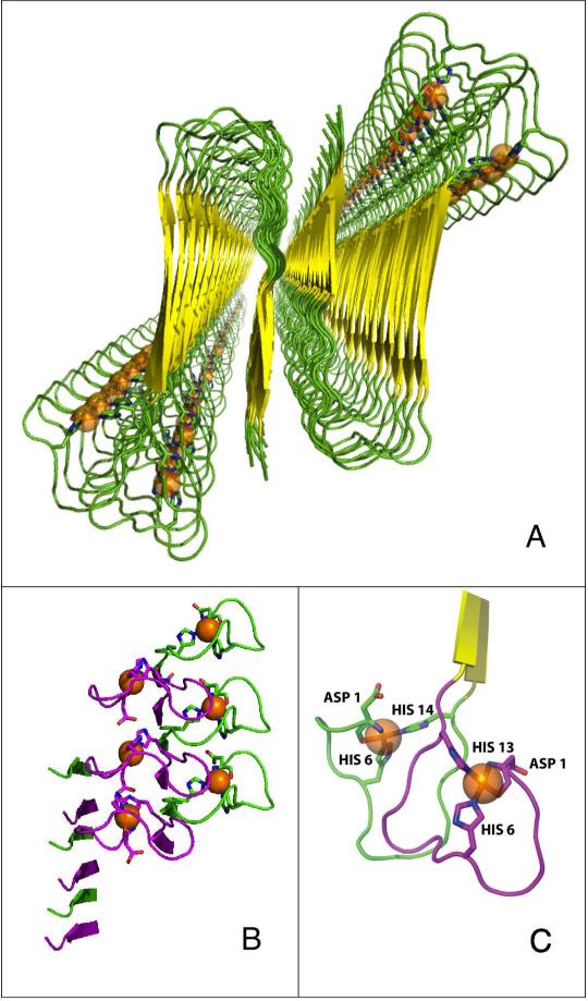
Figure 6.
Model for the coordination of Cu2+ in the fibrillar Aβ(1-40) peptide. The model is based on the quaternary structure for the ordered residues 9-40 of Aβ(1-40), which was determined by SS-NMR3 (Protein Data Bank ID, 2LMN). (A) Protein secondary structure cartoon, showing global view down the fibril axis. The Cu2+ ions are represented as orange spheres. (B) Side-on view of the N-terminal region, showing the patterning of Cu2+ sites along the fibril axis. Purple and green loops represent His6/His13 and His6/His14 sites, respectively. (C) Local Cu2+ coordination site geometry.

