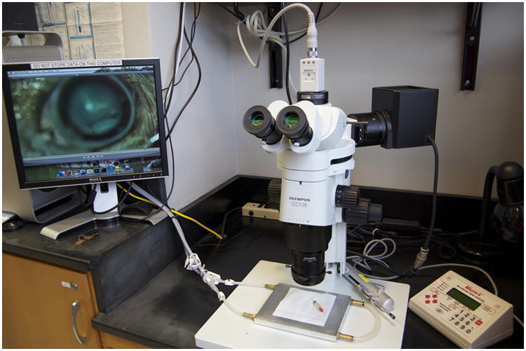
Figure 2.

Injection and microscope setup. A conventional dissecting microscope is used with an epifluorescence halogen light source. A video camera is mounted to the microscope. A nanoliter injection system from WPI is employed. An air filter to the right suppresses dust and air currents at the injection station. A computer for control of the video camera and for video editing is partially pictured to the left. A homemade aluminum stage warmer is shown on the stage. Temperature is controlled with a Lauda circulating water bath. During surgery, the aluminum block is covered with a small rectangle of fresh spill paper.
