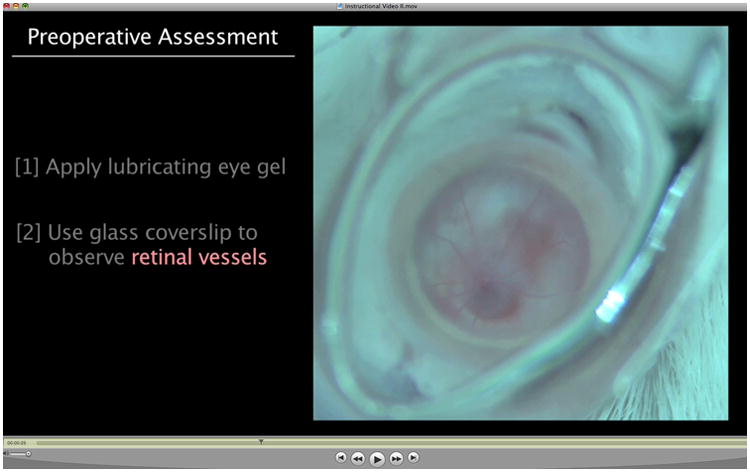
Figure 5.

Video of subretinal injection by transcorneal route. This video was created on an Olympus dissecting microscope equipped with a ring light and an HD video camera. Double-click on the image to play the video. The orange color upon subretinal injection comes from the fluorescence of quantum dots, which demarcate the extent of the subretinal bleb. The slide bar at the bottom of the Quicktime movie can be used to manually control the flow of the movie. The caption and figure image are from Johnson and coworkers (1), reprinted with permission.
