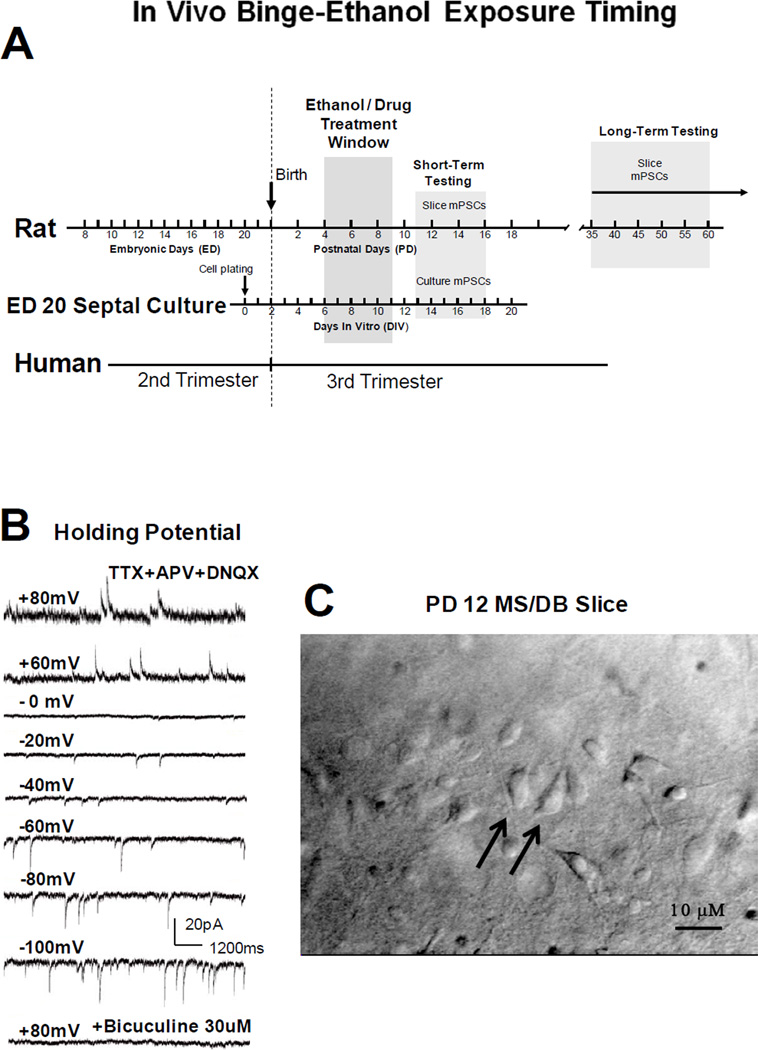
Figure 1. Schematic of binge-like ethanol intoxication timing, voltage-dependent whole cell GABAAR mPSC recordings, and a representative MS/DB rat brain slice.
(A) Relative human and rat brain development based on the ‘brain growth spurt’ concept of Dobbing and Sands [15]. Rats were treated during brain development equivalent to human 3rd trimester and when septo-hippocampal pathway formation is underway. GABAAR mPSCs were recorded after ‘binge ethanol’ in vivo (ethanol on PD 4–9, then slice recording PD 11–16). (B) GABAAR mPSCs reverse near 0 mV and are blocked by bicuculline as expected for a GABAAR-mediated chloride conductance under our recording conditions. (C) PD 12 coronal slice showing diagonal band neurons (40X water immersion, differential interference contrast optics, Olympus BX50WI microscope). Larger, bipolar neurons are indicated by arrows.

