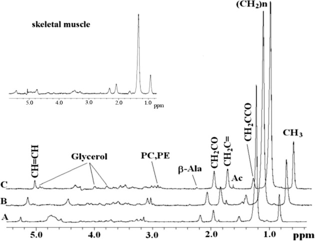
Figure 2.

In vivo 1D HRMAS 1H CPMG spectra of: (A) young wt injured, (B) old wt injured, and (C) young imd injured flies. Lipid components: CH3 (0.89 ppm), (CH2)n (1.33 ppm), CH2C-CO (1.58 ppm), acetate (Ac, 1.92 ppm), CH2C═C (2.02 ppm), CH2C═O (2.24 ppm), ß-alanine (ß-Ala, 2.55 ppm), phosphocholine (PC, 3.22 ppm), and phosphoethanolamine (PE, 3.22 ppm) glycerol (4.10, 4.30 ppm 1,3-CH; 5.22 ppm 2-CH2), CH═CH (5.33 ppm). The spectra in the insert are from the thorax of dissected flies and thus represent primarily skeletal muscle; note their similarity to spectra for whole flies. Shown spectra were normalized to TSP at each echo time and therefore do not exhibit T2 decay.
