
Fig. 6.
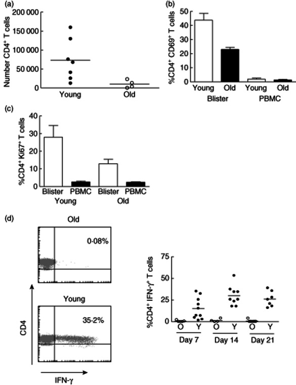
Reduced clinical responses to Mantoux test (MT) in old individuals are associated with reduced T cell accumulation and activation in the skin. (a) Skin suction blisters were raised on day 7 following MT induction in young and old individuals. Absolute number of CD4+ T cells in each blister was calculated using fluorescence activated cell sorter (FACS) analysis using Truecount tubes (n = 7 young and n = 4 old). (b) Cells isolated from skin suction blisters and peripheral blood on day 7 after the MT induction were stained with antibodies for CD3, CD4 and CD69 immediately ex vivo and analysed by flow cytometry. Graph shows the mean ± standard error of the mean of the percentage of CD4+ T cells expressing CD69 in blisters and peripheral blood mononuclear cells (PBMC) (Mann–Whitney U-test P = 0·0357; n = 4 young, 5 old). (c) Blister cells and PBMCs isolated on day 7 following MT were stained with antibodies for CD4 and Ki67 in order to identify CD4+Ki67+ T lymphocytes by flow cytometry (Mann–Whitney U-test P = 0·095; n = 5, mean ± standard error of the mean is shown). (d) Blister cells were collected at different times following MT induction and stimulated with purified protein derivative (PPD) ex vivo for 15 h in the presence of brefeldin. CD4+ T cells were then examined for intracellular interferon (IFN)-γ expression by flow cytometry. Representative FACS plots are shown on the left. The percentage of CD4+ T cells producing IFN-γ at different time-points is shown in the graph (horizontal lines represent the mean).