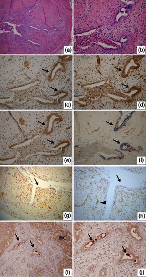
Fig. 1.

Histology and immunohistochemistry for interleukin (IL)-32, Toll-like receptor (TLR)-3 and caspase 1 in biliary atresia (BA). (a,b) Transverse sections of biliary remnants. Damaged extrahepatic bile ducts line inconsistently by desquamated columnar epithelium and surrounding fibroplasia with an inflammatory cell infiltrate; (b) a higher magnification of (a). Haematoxylin and eosin (H&E) staining. Original magnification (a) ×100 and (b) ×400. Immunohistochemistry for IL-32 (c), TLR-3 (d) and caspase 1 (e). The strong expression of IL-32, TLR-3 and caspase 1 was found in biliary epithelial cells (arrows) of damage bile ducts. Original magnification ×400. (f) Double immunohistochemistry for CK19 and IL-32 highlighted the CK19-positive bile ducts (blue) clearly expressed IL-32 (brown) (arrows). Original magnification ×400. (g,h) Immunohistochemistry for IL-32. Undamaged extrahepatic bile duct located at the resected margin in BA. IL-32-positive neovascular structures (arrowhead) were found, but undamaged biliary epithelium lacked IL-32 expression (arrows); (h) is higher magnification of (g). Original magnification (g) ×200 and (h) ×400. (i,j) Immunohistochemistry for IL-32 using wedge liver specimens of BA. Interlobular bile ducts (arrows in i) and hepatocytes (HC in i) expressed IL-32. Moreover, condensed bile in dilated bile ducts was also strongly positive for IL-32 (arrows in j); (j) is a higher magnification of (i). Original magnification (e) ×200 and (f) ×400.
