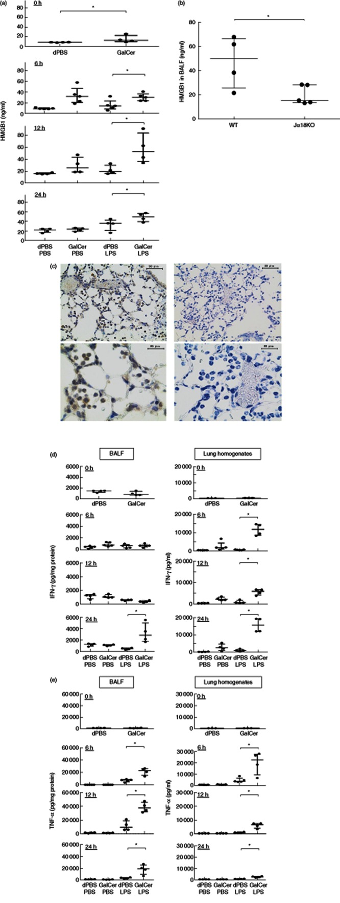
Fig. 1.
High mobility group box 1 (HMGB1) and cytokine production in the lungs of severe acute respiratory distress syndrome (ARDS) mice. Mice received intratracheal administration of lipolysaccharide (LPS) or phosphate-buffered saline (PBS) 24 h after administration of α-galactosylceramide (α-GalCer) or 0·4% dimethylsulphoxide (DMSO)-containing phosphate-buffered saline (dPBS) via the same route. (a) The HMGB1 concentration in the bronchoalveolar lavage (BAL) fluids was measured at 0, 6, 12 and 24 h after treatment with LPS or PBS. The data are shown as the median and interquartiles and each dot represents an individual mouse. (b) Jα18KO and wild-type mice received intratracheal administration of LPS 24 h after administration of α-GalCer via the same route. The HMGB1 concentration in the BAL fluids was measured 12 h after treatment with LPS. The data are shown as the median and interquartiles and each dot represents an individual mouse. *P < 0·05. (c) Mice received intratracheal administration of LPS 24 h after administration of α-GalCer via the same route. The lung sections, prepared 12 h after LPS treatment, were stained with anti-HMGB1 antibody (left) or control chicken immunoglobulin (Ig)Y (right). Representative pictures are shown at magnifications of ×400 (top) and ×1000 (bottom). (d,e) Mice received intratracheal administration of LPS or PBS 24 h after administration of α-GalCer or dPBS via the same route. The interferon (IFN)-γ (d) and tumour necrosis factor (TNF)-α (e) concentrations in the BAL fluids and lung homogenates were measured at 0, 6, 12 and 24 h after treatment with LPS or PBS. The measurements in BAL fluids are normalized to the respective protein content. The data are shown as the median and interquartiles and each dot represents an individual mouse. *P < 0·05.

