
Fig. 5.
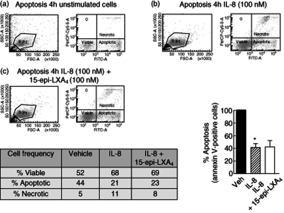
Measurement of interleukin (IL)-8-induced neutrophil survival by annexin V staining: effect of 15-epi-lipoxin (LX)A4. Cell apoptosis was measured in unstimulated human neutrophils (a) stimulated with IL-8 (100 nM) for 4 h (b) or preincubated with 15-epi-LXA4 (100 nM) for 30 min before addition of IL-8 (100 nM) for 4 h (c). Apoptosis induction was measured by annexin V staining. Propidium iodide staining was included for necrotic cell detection. Total neutrophils were gathered (left panel) and the number of viable neutrophils (double-negative cells), apoptotic neutrophils (annexin V-positive cells) and necrotic neutrophils (IP-positive cells) was detected using the proper probes by flow cytometry. Percentage of viable, apoptotic and necrotic cells are shown in the insert table. The percentage of apoptotic neutrophils after IL-8 and IL-8 in addition to 15-epi-LXA4 treatment compared to vehicle is represented in the bottom right graph. 15-epi-LXA4 did not increase annexin V-positive cells and did not reverse IL-8-induced neutrophil apoptosis arrest. *P < 0·05 IL-8 versus Vehicle.