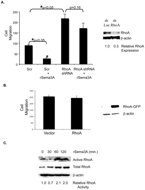
Figure 4. Sema3A stimulates RhoA activity and suppresses breast tumor cell migration in a RhoA-dependent manner.
A. Right panel: Equivalent amounts of protein from control shRNA-expressing (shLuc) and RhoA shRNA-expressing (shRhoA) MDA-MB-231 cells were subjected to SDS-PAGE and immunoblotted with RhoA or β–actin specific antibodies followed by the appropriate secondary antibody. Left panel: shLuciferase (shLuc)- and shRhoA-expressing MDA-MB-231 cells (80% confluent) were serum starved overnight prior to being incubated +/− rSema3A (100 ng/mL) for 40 min. The ability of these cells to migrate toward 5% FBS culture medium was assessed in a 4 hour assay. Data are presented as the mean number (+/− SD) of migrated cells from three wells (five fields/well). *p<0.05 in a Student’s t test. Similar data were obtained in three independent trials. B. MDA-MB-231 cells were transfected with pcDNA3-EGFP or pcDNA3-EGFP-RhoA-wild-type using Fugene HD (Roche, Indianapolis, IN) After selection in neomycin for 1 week, the migratory potential of these transfectants was assessed in transwells, as described for A. C. MDA-MB-231 cells were incubated +/− rSema3A for the indicated times, and RhoA activity was measured using Rhotekin RBD beads, which bind specifically to activated (GTP-bound) RhoA. These immunoprecipitates were subjected to SDS-PAGE, and immunoblotted with a RhoA antibody, followed by the appropriate species secondary antibody. As a loading control, lysates from these samples were also immunoblotted with a RhoA or β–actin antibody. Similar data were obtained in three independent trials.

