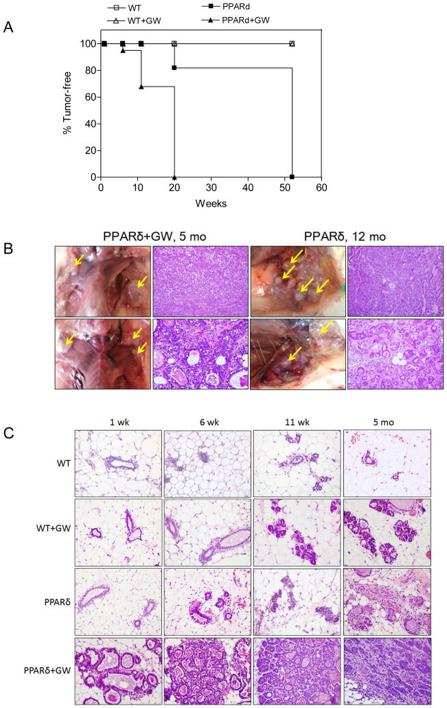
Figure 1. PPARδ activation promotes mammary neoplasia.
A, Tumor-free incidence in MMTV-PPARδ mice fed a standard or GW-supplemented diet. Tumor formation differed significantly between untreated or GW501516-treated PPARδ mice (PPARd+GW) vs. untreated or GW-treated wild-type mice (WT+GW) (P<0.0001 by the Chi2 test). GW-treated PPARδ mice (PPARd+GW) developed tumors more rapidly vs. untreated PPARδ mice (P<0.0001 by the Chi2 test). Each time point represents tumor incidence as determined by histological examination post-mortem. The number (N) of mice per group at each time point was: WT (N=8), WT+GW (N=8), PPARd (N=6), PPARd+GW (N=7). B, Tumor formation in situ. PPARδ mice fed a standard or GW-supplemented diet presented with multifocal moderate to well-differentiated infiltrating ductal carcinomas at 12 months or five months, respectively. Shown are representative mice from each group. H&E, magnification, 400X. C, H&E stained tissue. GW-treated wild-type (WT) exhibited ductal dilitation and secretory changes after six weeks, and increased ductal branching after 11 weeks to five months. PPARδ mice presented with increased ductal branching after six weeks, lobular dysplasia after 11 weeks and areas of neoplasia after five months. GW-treated PPARδ mice exhibited ductal dilatation with lipid droplets and proteinaceous secretions after one week, atypical lobular and ductal hyperplasia after six weeks, atypical ductal dysplasia bordering on ductal carcinoma in situ after 11 weeks and infiltrating ductal carcinomas after 5 months. Magnification, 400X.

