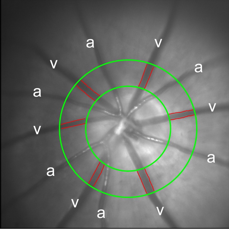
Figure 2.
An example of a red-free retinal image displaying six major retinal arteries (a) and veins (v) in a rat under normoxia. The outlined edges of the retinal veins (red lines) were identified from multiple-diameter measurements over a fixed vessel span at a defined distance from the center of the optic nerve head, as delineated by green circles.

