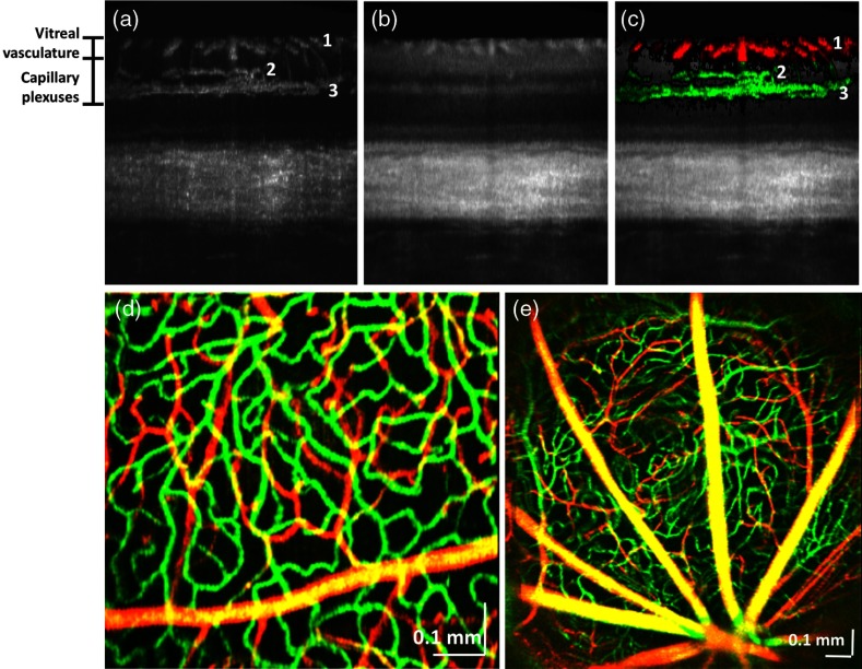
Fig. 1.
Visualization of the rat retinal vasculature with OCT angiography. (a) OCT angiogram in cross-section, showing vasculature in the nerve fiber layer (1), inner plexiform layer (2), and outer plexiform layer (3). (b) Corresponding cross-sectional OCT intensity image. (c) Overlay of OCT angiogram (coloring the capillary plexus green and the vitreal vessels red) with the standard intensity image, showing supplying and draining vessels descending or ascending through the ganglion cell layer to both the inner and outer plexiform layers. (d) En face color image of vasculature in the peripheral retina, showing that the angiographic technique depicts subtle differences in vessel diameter. (e) Wide-field image of the vasculature near the optic nerve head.

