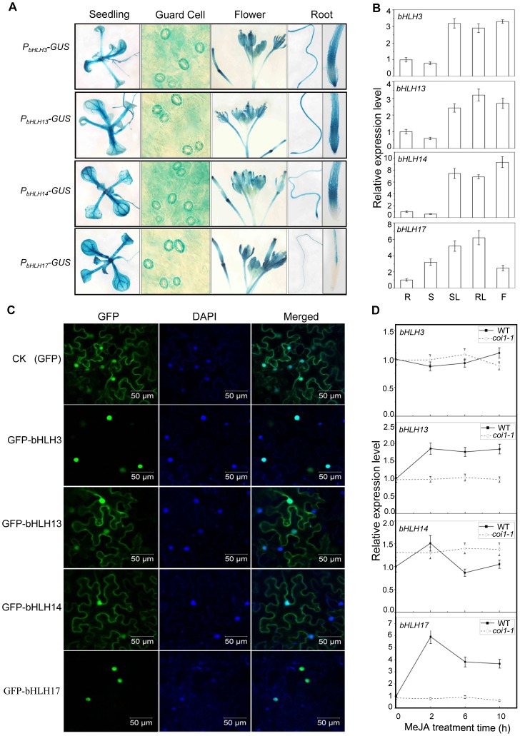
Figure 2. Expression patterns and subcellular localizations of bHLH3, bHLH13, bHLH14 and bHLH17.
(A) GUS reporter gene was fused with the promoters of the four bHLH factors respectively to generate Arabidopsis transgenic plants (PbHLH3-GUS, PbHLH13-GUS, PbHLH14-GUS and PbHLH17-GUS). Histochemical GUS activity was detected in various tissues of transgenic seedlings. (B) Quantitative real-time PCR analysis of relative expression levels of bHLH3, bHLH13, bHLH14 and bHLH17 in root (R), stem (S), rosette leaf (RL), stem leaf (SL) and flower (F). ACTIN8 was used as the internal control. Error bars represent SE (n = 3). (C) Subcellular localization of bHLH3, bHLH13, bHLH14 and bHLH17 in epidermal cells of N. benthamiana leaves. Constructs indicated on the left were infiltrated in leaves of N. benthamiana. GFP fluorescence was detected 50 hours after infiltration. The nuclei were indicated by DAPI staining. (D) Quantitative real-time PCR analysis of bHLH3, bHLH13, bHLH14 and bHLH17 in 11-day-old WT and coi1-1 seedlings treated with 100 µM methyl-jasmonate (MeJA) for 0, 2, 6, and 10 hours. ACTIN8 was used as the internal control. Error bars represent SE (n = 3).

