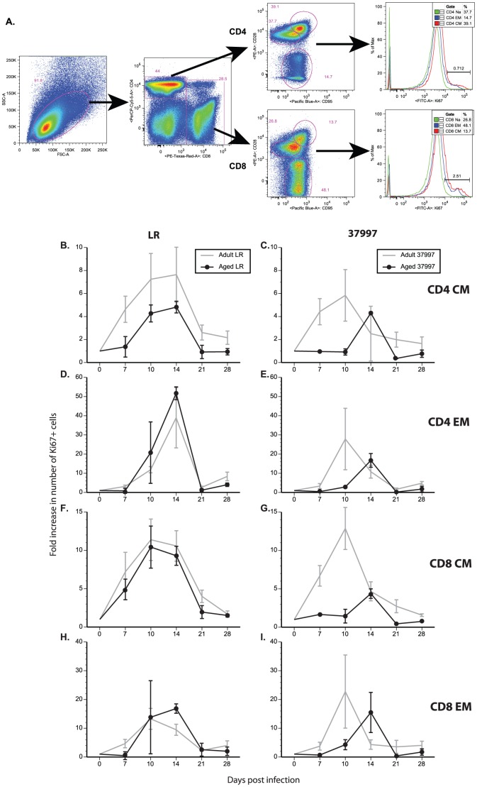
Figure 3. Aged Rhesus macaques have lower T cell responses following CHIKV infection.
T-cell proliferative burst was measured following CHIKV infection. PBMC were stained with antibodies directed against CD4, CD8, CD28, CD95 and Ki67 (as shown in panel A). Fold increase in number of Ki67+ cells was calculated for each time point. Cell populations were subdivided: B & C) CD4+ Central memory (CM) T-cells; D & E) CD4+ Effector memory (EM) T-cells; F & G) CD8+ CM; H & I) CD8+ EM for animals infected with CHIKV-LR (panels B, D, F, H) or CHIKV-37997 (panels C, E, G, I). N = 6 for adult and N = 2 for aged animals.

