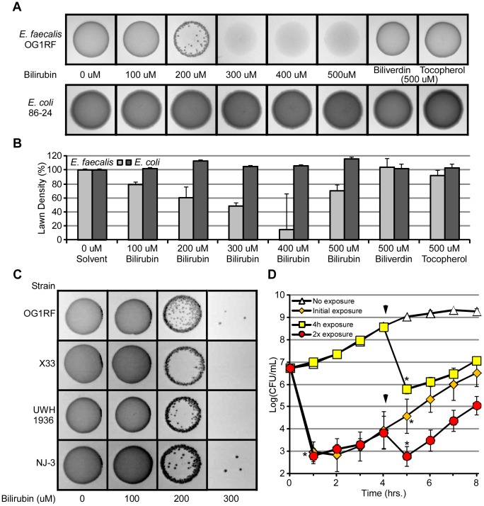
Figure 4. Bilirubin decreases the viability of E. faecalis.
(A) An equal number of CFUs of E. faecalis or E. coli (O157:H7 86-24) were mixed with increasing amounts of bilirubin (0, 100, 200, 300, 400, and 500 µM), biliverdin (500 µM), or α-tocopherol (500 µM), before spotting onto LB agar. Plates were grown at 37°C overnight. (B) Densitometry of bacterial lawns was measured using ImageJ software. (C) E. faecalis strains OG1RF (oral origin), X33 (fecal origin), UWH 1936 (blood origin), and NJ-3 (peritoneal fluid origin) were exposed to increasing concentrations of bilirubin (100, 200, or 300 µM) prior to spotting onto an LB-agar plate. Bacterial growth was captured by imaging the plates, and images were modified by adjusting brightness and contrast to best display colony formation (darker areas of image). (D) Growth of E. faecalis with bilirubin (yellow squares) or without bilirubin (white diamonds) was quantified by CFU plating. Bilirubin was titrated into E. faecalis cultures initially with bilirubin (red squares) and initially without bilirubin (orange squares) after 4 hours (marked by arrows). Error bars represent ± one standard deviation, n = 3, and (*) denotes a significant (P≤0.05) difference between treated samples and solvent-treated samples.

