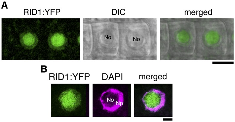
Figure 6.
Subcellular Localization of RID1:YFP.
Fluorescence signals of RID1:YFP in the RAM. The fluorescence images of RID1:YFP signal were merged with the differential interference contrast (DIC) image (A) and with the fluorescence image of DAPI staining (B). No, nucleoli region; Np, nucleoplasmic region. Bars = 10 µm in (A) and 5 µm in (B).

