Figure 7.
GUS Analysis of RID1 Promoter Activity.
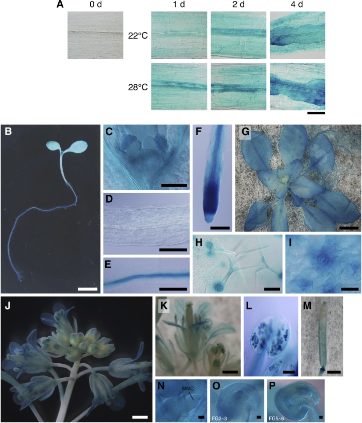
(A) ProRID:GUS expression patterns during callus initiation. Hypocotyl explants of 12-d-old RID:GUS seedlings were cultured on CIM for the indicated times at 22 or 28°C and then subjected to histochemical detection of GUS activity. Bar = 100 µm.
(B) to (F) ProRID1:GUS expression patterns in 6-d-old seedlings. (C) to (F) show magnified images of the shoot apex, hypocotyl, root stele, and root tip tissues, respectively. Bars = 5 mm in (B) and 100 µm (C) to (F).
(G) to (I) RID1:GUS expression patterns in 24-d-old transgenic plants. Panels show the macroscopy image (G) and higher magnifications ([H] and [I]) of the leaf surface. Bars = 5 mm in (G), 100 µm in (H), and 25 µm in (I).
(J) to (P) ProRID1:GUS expression patterns in the reproductive phase. Panels show an inflorescence (J), opening flower (K), anther containing mature pollens (L), pistil after fertilization (M), megaspore mother cell–containing ovule (N), ovule at stage FG2/3 (O), and ovule at stage FG5/6 (P). MMC, megaspore mother cell. Bars = 1 mm in (J) and (M), 0.5 mm in (K), 100 µm in (L), and 10 µm in (N) to (P).

