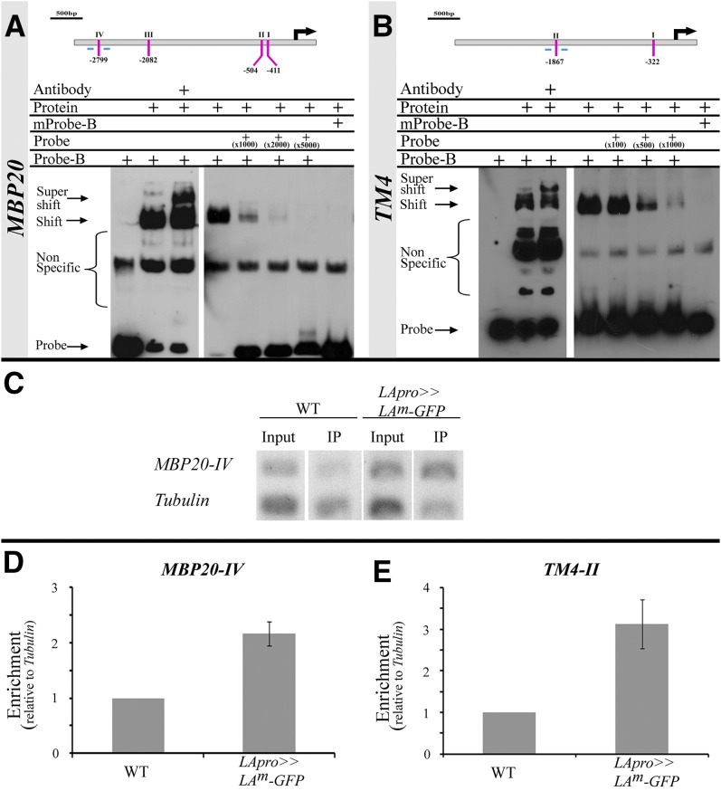
Figure 4.
LA Binds to the MBP20 and TM4 Promoters.
(A) and (B) Top: Schematic diagrams of the MBP20 (A) and TM4 (B) promoters and potential core TCP binding sites (GGNCC, indicated with lines and roman numerals). Black arrows indicate the translation start sites. Bottom: EMSA performed with biotin-labeled probes (Probe-B) and recombinant LA protein fused to MBP (Protein). The components included in each reaction are indicated above each lane. Probe, unlabeled probe (folds of the amount of labeled probe indicated); mProbe and mProbe-B, unlabeled and labeled mutated probe (…GGNaCt…), respectively; Antibody, antibodies against MBP.
(C) to (E) PCR and quantitative PCR analyses of a ChIP assay, performed with wild-type plants (WT) or plants expressing a LAm-GFP fusion under the control of the LA promoter (LApro>>LAm-GFP) and anti-GFP antibodies.
(C) PCRs were performed with specific primers for MBP20-IV (lines below the gene diagrams in [A]) and Tubulin. Input, nonimmunoprecipitated samples; IP, samples after ChIP.
(D) and (E) Quantitative PCR reactions were performed with specific primers for MBP20-IV (D) or TM4-II (E) (lines below the gene diagrams in [A] and [B], respectively) or Tubulin. Shown are averages (±se) of fold enrichment, compared with the wild type (n = two technical and three biological repeats in [D] and three biological repeats in [E]).
[See online article for color version of this figure.]

