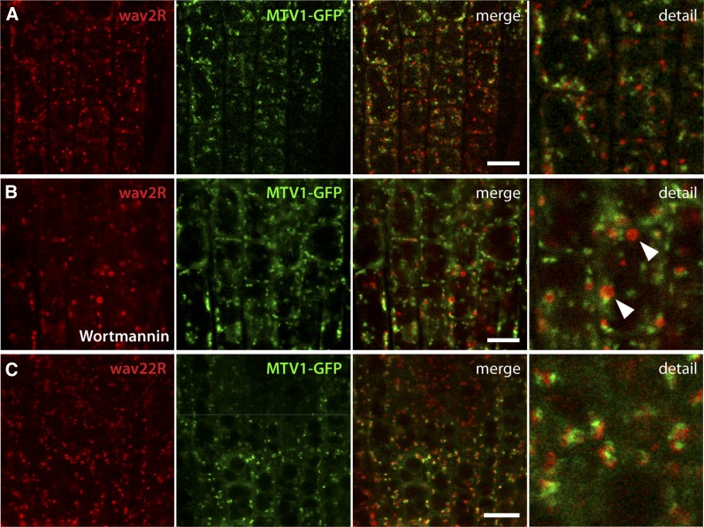
Figure 4.
MTV1 Is Separate from the PVC and the cis-Golgi.
(A) Live-cell imaging of root epidermal cells harboring the PVC marker wave2R (ARA7) and MTV1-GFP reveals no overlap.
(B) Incubation with 30 µmol wortmannin for 1 h caused formation of wave2R-labeled ring-like structures (arrowheads), and no colocalization with MTV1 was observed.
(C) The cis-Golgi marker wave22R (SYP32) did not colocalize with MTV1, but signals were typically found in close vicinity.
Bars = 20 µm.

