Figure 4.
Spatiotemporal Accumulation of ClpS1 and Interaction between ClpS1 and ClpC Proteins.
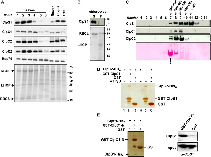
(A) Arabidopsis plants were grown on soil under continuous light. Leaves from the two outer rows of the rosette were harvested after 1 to 6 weeks. Flowers, siliques, and stems were collected after 6 weeks. Total proteins were extracted and analyzed by immunoblotting using anti-ClpS1, ClpC1/C2/R2, and cpHsp70 antibodies. Each lane contains 20 μg proteins, and the Ponceau-stained blot is shown as the loading control. Loss of RBCL during senescence (4 to 6 weeks) can be observed from the stained blot.
(B) ClpS1 is exclusively located in the stroma and is absent in the chloroplast membranes. To determine the intraplastid location of ClpS1, chloroplasts were isolated from soil-grown wild-type. The stromal (S) and membrane fractions (P) were separated by centrifugation and analyzed by SDS-PAGE and immunoblotting. The filter was also stained with Ponceau S (bottom panel).
(C) In vivo sizes of native ClpS1 and ClpC1/2 in the chloroplast stroma. Chloroplasts were isolated from soil-grown wild-type plants at leaf stage 1.07-1.08. The stromal proteins were prepared in the presence of ATPγS and separated using a Superose (gel filtration) column. The eluates were collected and pooled into 14 fractions. Proteins in each fraction were TCA precipitated, and equal volumes were analyzed by immunoblotting with antibodies against ClpS1, ClpC1, and ClpC2. The blot was also stained by Ponceau S, showing RBCL eluting as part of the 550-kD holocomplex (marked with an asterisk).
(D) Direct interaction of ClpS1 with ClpC2. ClpC2-His6 protein was incubated with or without ATPγS and subsequently combined with GST-ClpS1 or ClpS. Proteins were bound to the GST affinity resin and then eluted with reduced glutathione. Eluates were analyzed by SDS-PAGE and silver staining.
(E) Recombinant his6-tagged ClpS1 was incubated with recombinant GST or the N-domain of ClpC1 fused to GST. Proteins were bound to the GST affinity resin and then eluted with laemmli buffer. Eluates were analyzed by SDS-PAGE and silver staining, and ClpS1 was also detected by immunoblot with anti-ClpS1 antiserum.
[See online article for color version of this figure.]

